X-ray Imaging
GALLERY
X-ray Imaging
Exposure time :60ms/projection
Number of projections :1800 (Total imaging time approx. 5 min)
Sample-detector distance :150mm
X-ray energy :White X-rays (using 20mm Al absorber)
Pixel size : 20 μm
Exposure time :60ms/projection
Number of projections :1800 (Total imaging time approx. 5 min)
Sample-detector distance :150mm
X-ray energy :White X-rays (using 20mm Al absorber)
Pixel size :20 μm
Exposure time :60ms/projection
Number of projections :1800 (Total imaging time approx. 5 min)
Sample-detector distance :150mm
X-ray energy :White X-rays (using 20mm Al absorber)
Pixel size :5 μm
Exposure time :60ms/projection
Number of projections :1800 (Total imaging time approx. 5 min)
Sample-detector distance :150mm
X-ray energy :White X-rays (using 20mm Al absorber)
Pixel size :5 μm
Exposure time :60ms/projection
Number of projections :1800 (Total imaging time approx. 5 min)
Sample-detector distance :150mm
X-ray energy :White X-rays (using 20mm Al absorber)
Pixel size :5 μm
Exposure time :100ms/projection
Number of projections :1800
X-ray energy :15keV
Pixel size :0.65 μm
Exposure time :100ms/projection
Number of projections :1800
X-ray energy :15keV
Pixel size :0.65 μm
Exposure time :100ms/projection
Number of projections :1800
X-ray energy :15keV
Pixel size :0.65 μm
Exposure time:100ms/projection
Number of projections:1800
X-ray energy:9 keV
Pixel size:0.65 μm
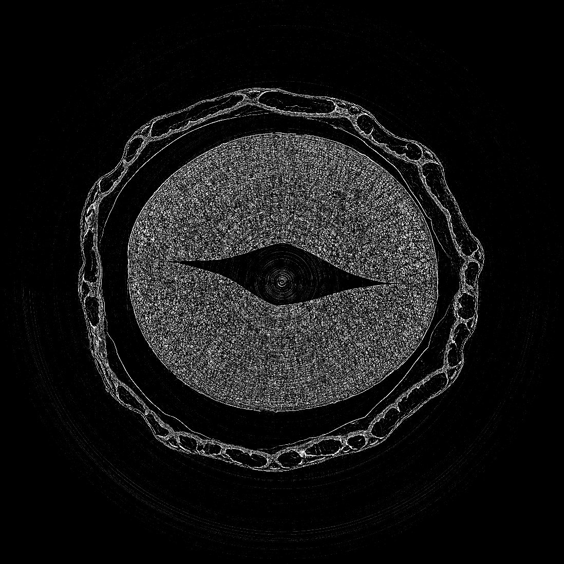
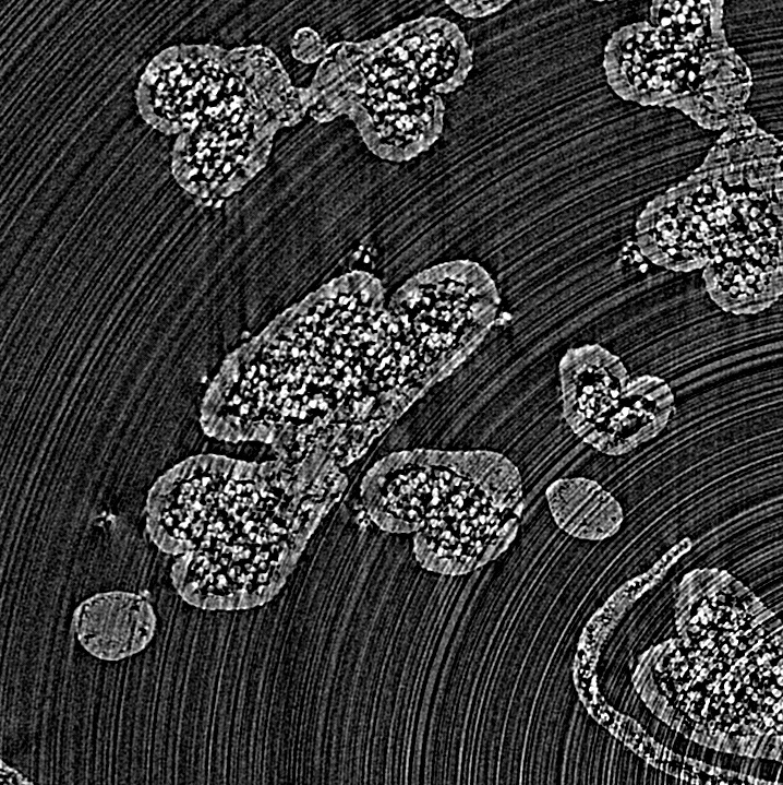
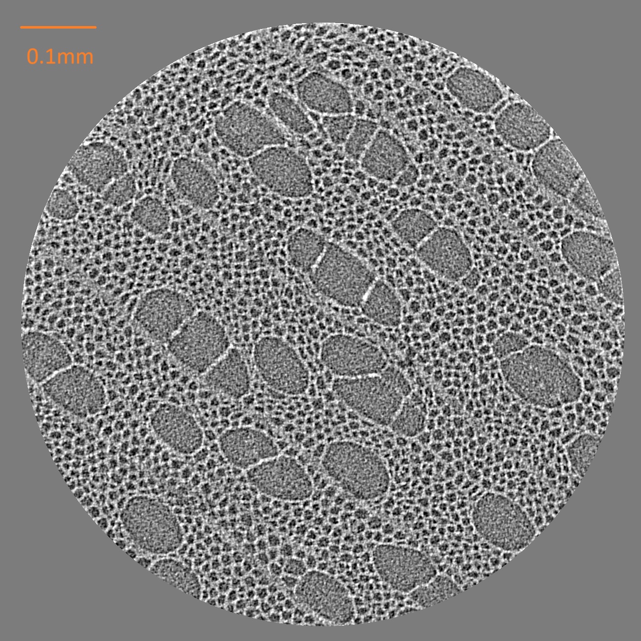
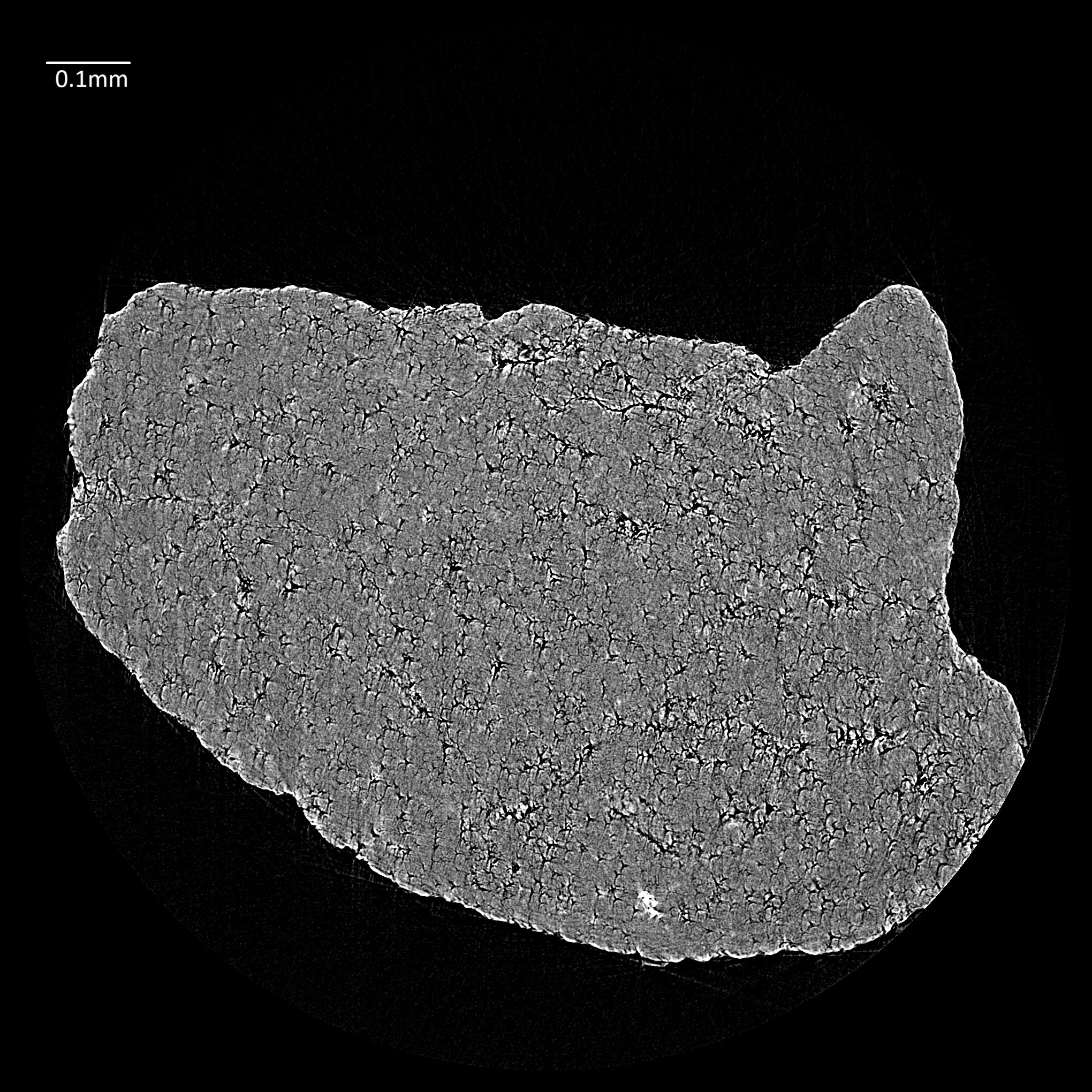
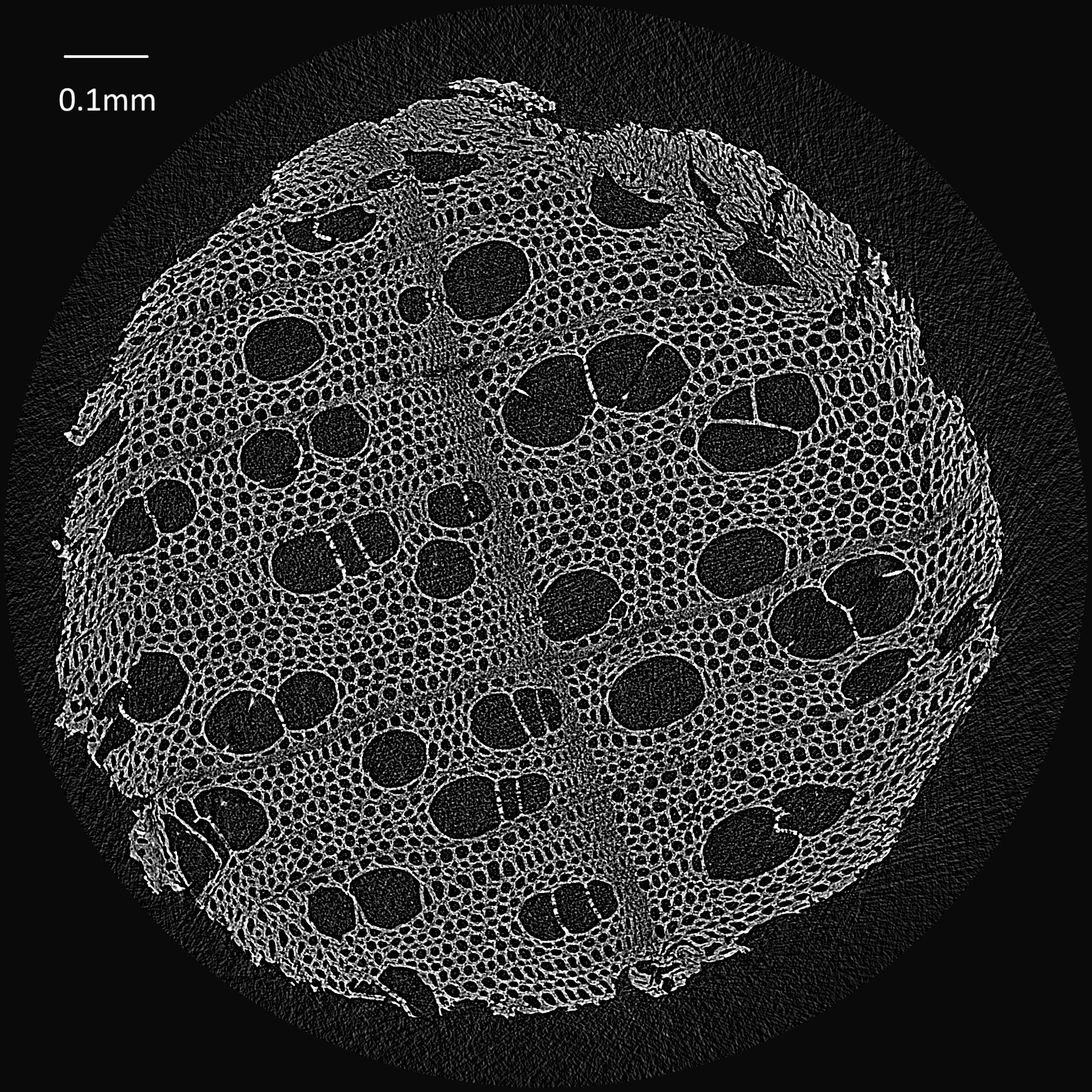
Looking at the image, many large black circular to elliptical pores and numerous fine honeycomb-like pores filling the surroundings can be seen in the cross-section of the toothpick. Biologically, this is interpreted as wood tissue, specifically the secondary xylem of a plant. The large pores can be interpreted as vessels responsible for water conduction, the fine pores as the cell lumens of wood fibers or parenchyma cells, and the white network-like parts primarily as cell walls containing lignin.
Particularly important is the presence of many large vessel-like pores. This suggests that the toothpick material is a wood tissue with vessels similar to hardwood, rather than a simple tracheid tissue typical of softwoods. However, the tree species cannot be identified from the image alone. Species identification requires a comparison of vessel arrangement, rays, parenchyma arrangement, and growth ring boundaries across multiple sections.
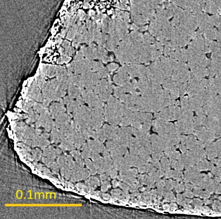
Furthermore, the fact that the vessels and cell lumens are open indicates that even though a toothpick appears to be a solid stick, its interior consists of a porous biological material. This porous structure is a remnant of the tissue that was involved in water and inorganic ion transport, mechanical support, and storage when the tree was alive. Even after becoming a toothpick, its cellular structure is preserved, and X-ray CT allows for its non-destructive observation.
The areas where the fine honeycomb structures are densely distributed are thought to be regions with many fiber tissues that provide the strength of the wood. On the other hand, regions with many large vessels have high porosity and are thought to have locally low density. Therefore, from this image, we can read not only that "pores are visible," but also how the tissues involved in water transport and those responsible for strength are spatially arranged.
At the outer periphery and some edges, some parts appear collapsed, peeled, or cracked. This may reflect damage during processing (cutting, polishing, drying) into a toothpick, or mechanical deformation as a thin piece of wood. While the cell wall structures of biological origin remain, it is appropriate to view this as observing both the internal damage and deformation as a processed wood material rather than just the natural wood itself.