
Monochromatic X-ray CT
(High-resolution CT)
TECHNICAL INFORMATION
Any unauthorized reproduction, copying, or alteration of information on this website is prohibited.
Latest news and schedules for beamline upgrades are available on this page. Please also check it for updates.
Basic Information
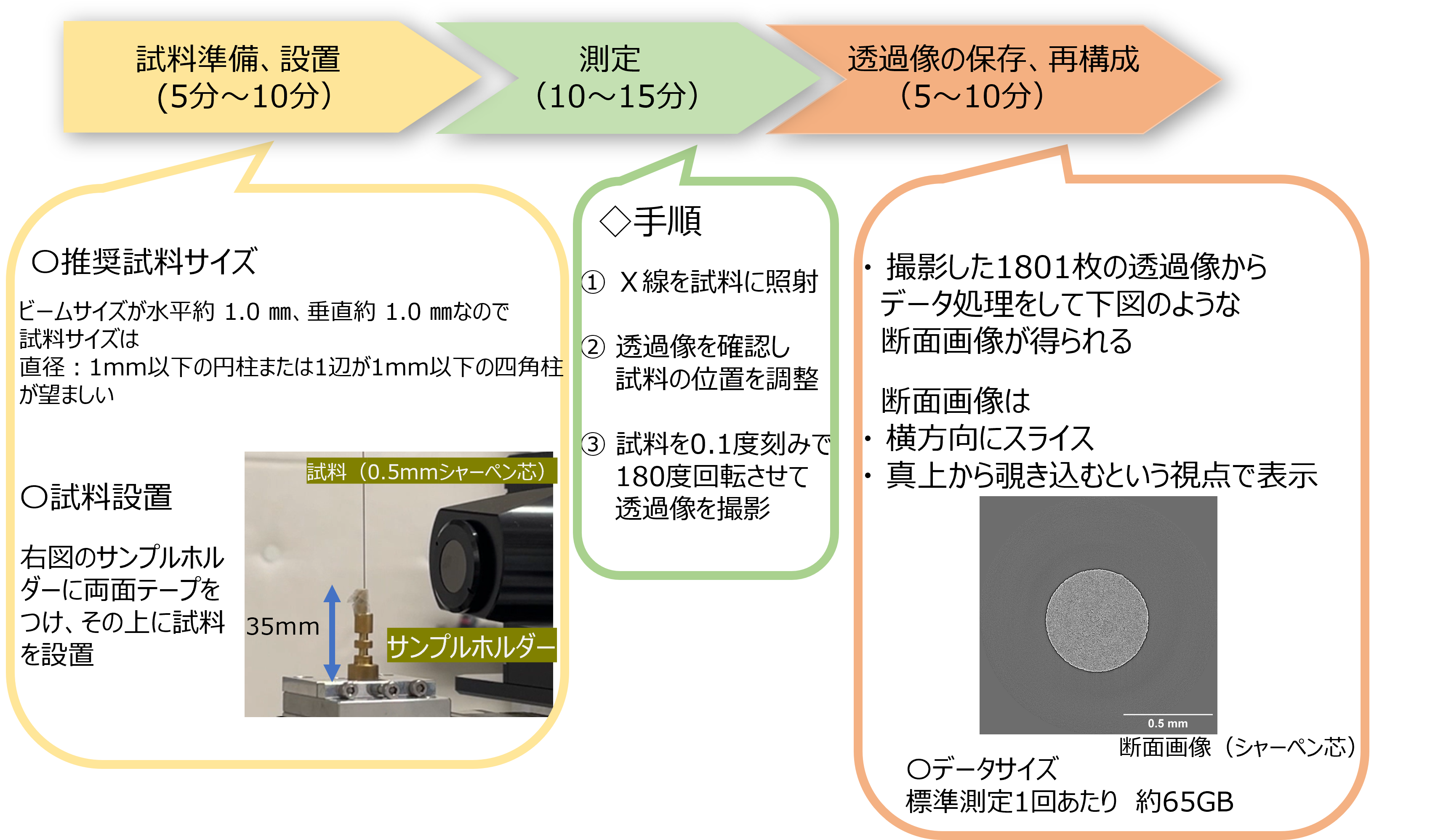
- Field of ViewApprox. 1 mm (horizontal) x 1 mm (vertical)
- Spatial ResolutionApprox. 1 μm
- Pixel Size0.65 μm
- Measurement TimeApprox. 10 minutes per sample
- Data Volume65 GB per sample (Please bring a USB storage of 2 TB or more)
-
Energy
Standard energy options:
• 8.33 keV
• 11.67 keV
• 15 keV
• 18.33 keV
You can change between these standard energy levels during your beamtime. The energy switch takes about 5 minutes.
However, if you require a different energy (for example, for a specific metal absorption edge), please consult with us at the time of reservation. - Sample SizeIdeally cylinder < 1 mm dia. or prism < 1 mm side. Length: 10 - 50 mm.
- Sample MountingFixing to a cylindrical holder (double-sided tape, clay, etc.)
- Measurement Method180° rotation in 0.1° increments (Reconstructed from 1800 projections)
- Data TransferSSD with transfer speed of 1000 MB/s or higher is recommended.
Layout
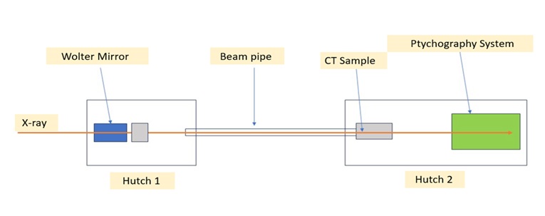
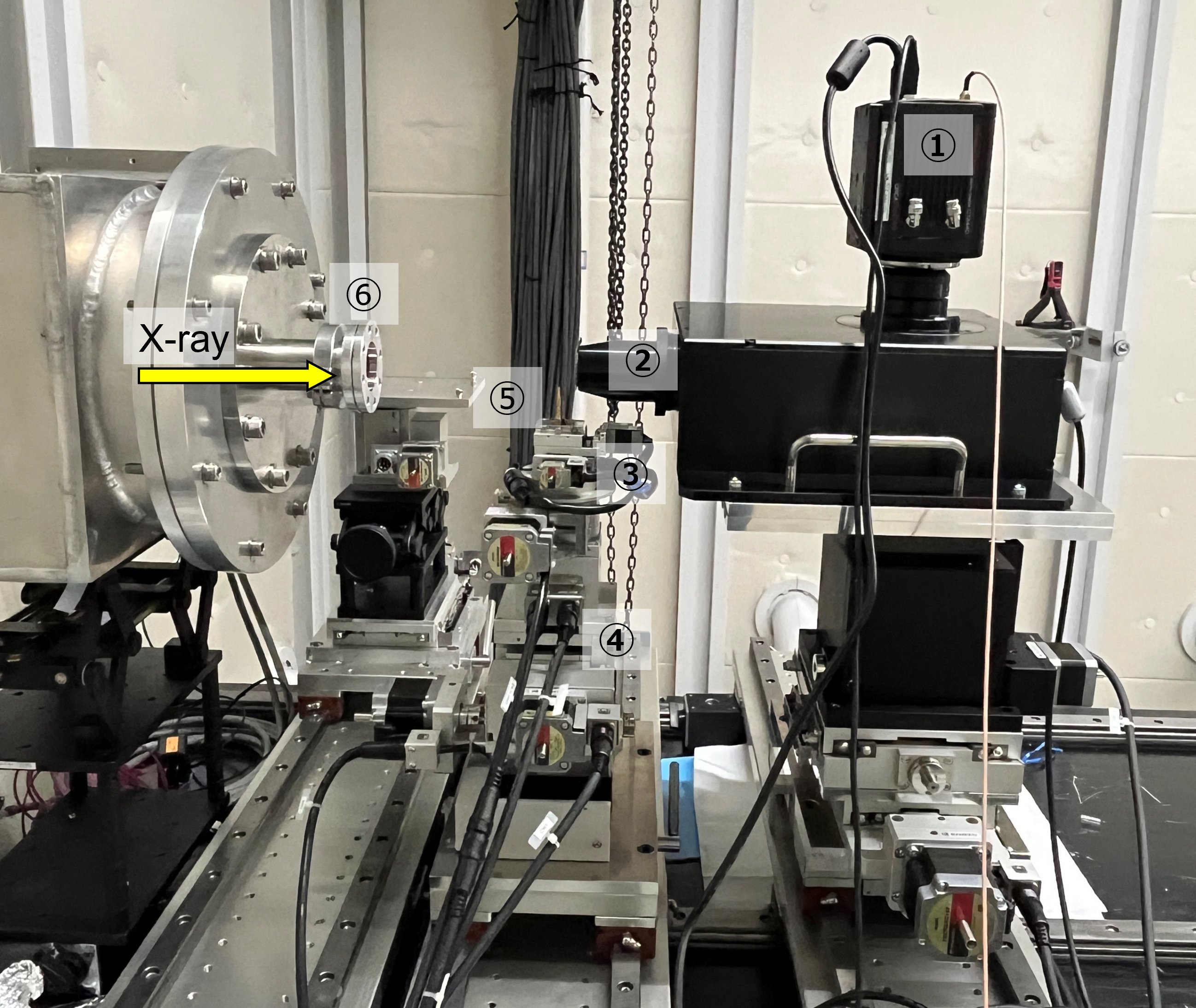
-
①
CMOS camera
Model: C13440-20CU (Hamamatsu Photonics) - ② Scintillator
-
③
X-Y Stage
Model: YA-04-R102-NNR (Kozu Seiki)
Travel Range: ±5 mm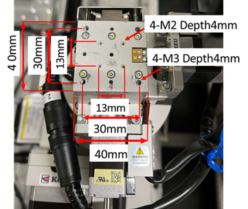
-
④
Z Stage
Model: ZA-10A-W202 (Kozu Seiki)
Travel Range: ±7 mm -
⑤
Sample Stage
A plate with a square hole (as shown in the figure below) is mounted to stage ③ using M3 screws.
The sample holder is placed into this square hole.
The beam irradiation position is 49 mm above the top surface of the stage.
The Z stage can move ±7 mm from that position.
If additional height is required, 5 mm or 10 mm spacers can be used.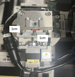
- ⑥ Kapton Window (Thickness: 75 μm)
Measurement Flow
Measurement Examples
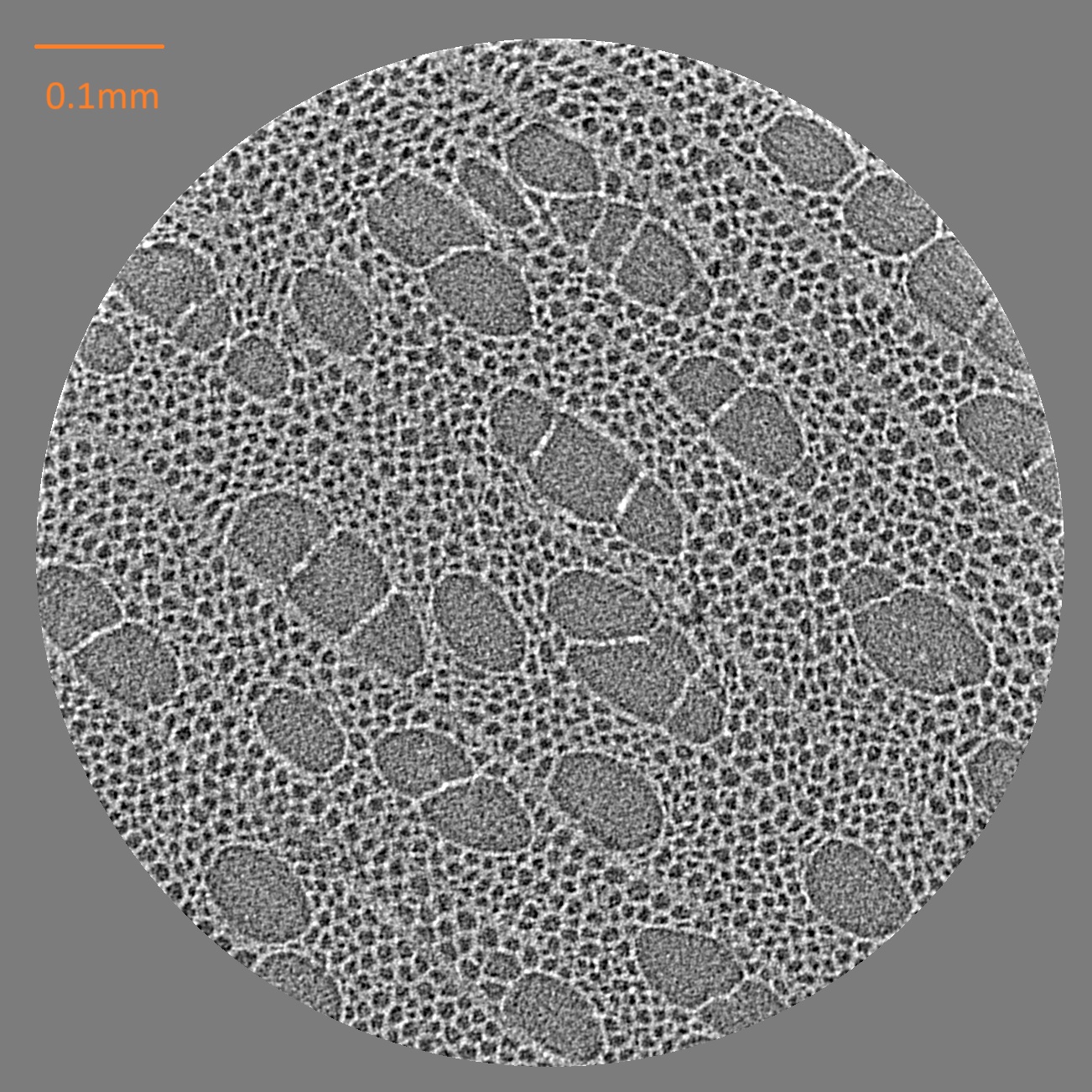
Toothpick (15 keV)
Exposure: 100 ms/projection, Projections: 1800, Pixel size: 0.65 μm
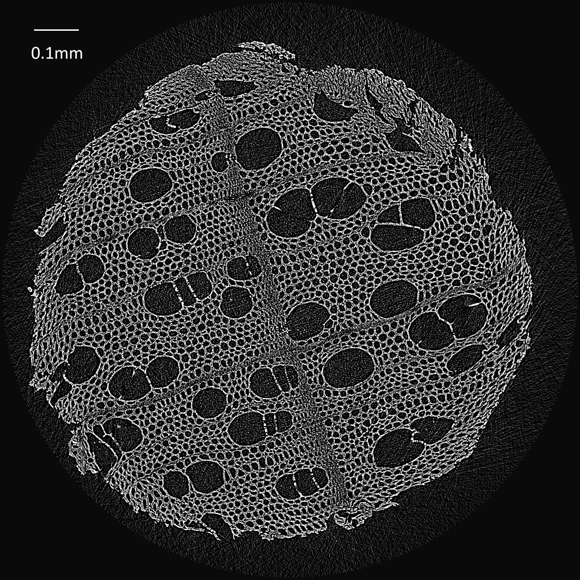
Toothpick (9 keV)
Exposure: 100 ms/projection, Projections: 1800, Pixel size: 0.65 μm
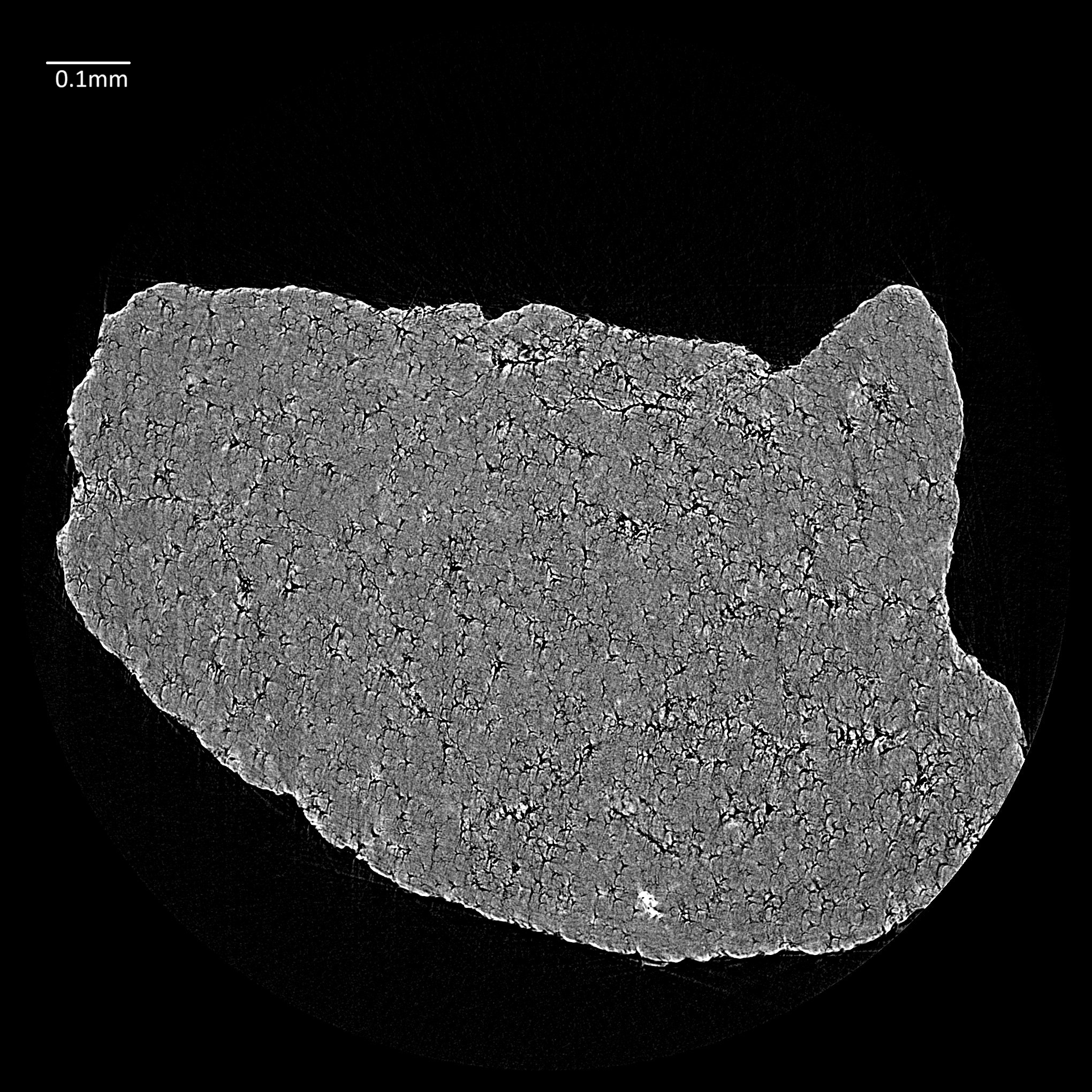
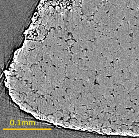
Sand Grain
Energy: 15 keV, Pixel size: 0.65 μm
Rice Fragment
Energy: 15 keV, Pixel size: 0.65 μm
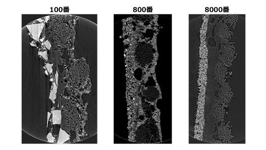
Sandpaper (15 keV)